Service Category: Heart

High Tibial Osteotomy

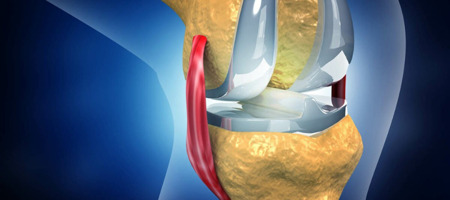
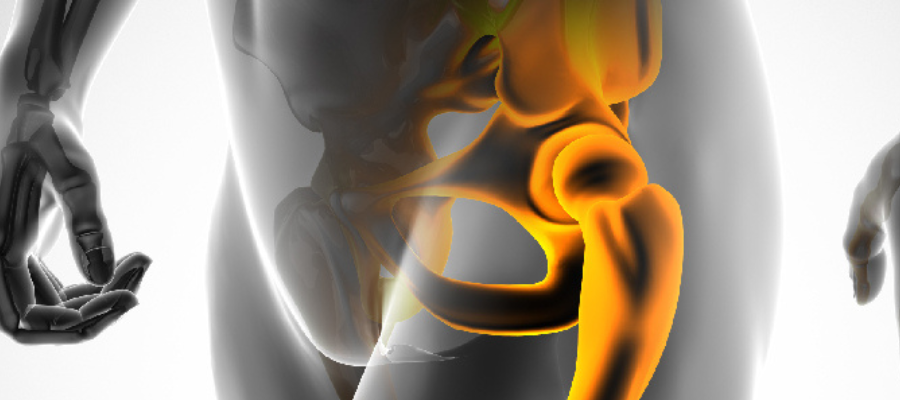
What is High Tibial Osteotomy? High tibial osteotomy (HTO) is a widely performed procedure to treat medial knee arthrosis. If one part of the joint is worn out, the angle of the leg bone can often be changed to shift stresses onto other areas that are not so worn. This procedure can produce years of pain relief, as an alternative to joint replacement, in appropriate patients. The ideal candidate for an HTO is a middle aged patient (45 to 65 years of age), with isolated medial osteoarthritis, with good range of motion and without ligamentous instability. HTO significantly affects a subsequent total joint replacement. Precise indication, preoperative planning, and operative technique selection are essential to achieve good results. Purpose There are two main reasons to perform high tibial osteotomy: For patients with medial compartment arthritis and a varus knee, its purpose is to provide years of relief prior to knee replacement. This is particularly important in patients too young to be optimal candidates for knee replacement. The other reason it is done is to correct malalignment in patients undergoing another procedure such as Carticel implantation. In these patients the HTO is performed to protect the cartilage replacement from failure due to excessive compressive forces on the graft. HTO techniques There are various HTO techniques including closing wedge osteotomy, opening wedge osteotomy, dome osteotomy, progressive callus distraction, and chevron osteotomy. Medial Opening Wedge Osteotomy- Medial opening wedge osteotomy is a relatively simple procedure that involves a single osteotomy and a few dissections. The technique does not necessitate either a fibular osteotomy that has been associated with neurovascular complications or bone resection of the lateral tibia. Lateral Closing Wedge Osteotomy- Lateral closing wedge osteotomy is effective for correction near maximal point of deformity. The technique allows rapid bone union due to the large contact surface of cancellous bone at the osteotomy site, early weight bearing and rehabilitation, and the use of quadriceps femoris muscle force. Other Techniques- Other HTO techniques include dome osteotomy, progressive callus distraction using an external fixator, and chevron osteotomy. Outcome For most patients, osteotomy is successful in relieving pain and delaying the progression of arthritis in the knee. It can allow a younger patient to lead a more active lifestyle for many years. Even though many patients will ultimately require a total knee replacement, an osteotomy can be an effective way to buy time until a replacement is required. Common FAQs How long does recovery take after High Tibial Osteotomy? Most patients begin walking with support (crutches) within the first few weeks, with partial weight-bearing initially and gradual full weight-bearing over time. Full recovery typically takes about 3–6 months, and return to sports or high-impact activities may take 4–6 months or longer depending on progress and physiotherapy. Will I be able to walk on my leg right after surgery? Walking usually starts early with support. Initially you may use crutches or a walker with limited weight-bearing. As healing progresses, your surgeon and physiotherapist will advise when you can increase weight-bearing and walk more normally. When can I return to work and daily activities? Return to work depends on your job and knee recovery. Many patients return to light or desk-based work within a few weeks, but physically demanding jobs or sports may take several months before being safe to resume. What are the common risks after High Tibial Osteotomy? Like any bone surgery, potential risks include infection, delayed bone healing or non-union, stiffness, irritation from hardware (plates/screws), nerve or blood vessel issues, and swelling. Regular follow-ups and good physiotherapy reduce these risks. Can I return to sports after HTO surgery? Yes, many patients are able to return to sport and active lifestyles. Most can resume low-impact activities by 3–6 months, and some return to higher-impact activities by 6–12 months depending on healing, age, fitness, and medical advice.

Fracture Fixation

What is Fracture Fixation? A fracture is a broken bone. It can range from a thin crack to a complete break. Bone can fracture crosswise, lengthwise, in several places, or into many pieces. Most fractures happen when a bone is impacted by more force or pressure than it can support. Most fractures are accompanied by intense pain when the initial injury occurs. It may become worse when you move or touch the injured area. In some cases, you may even pass out from the pain. You may also feel dizzy or chilled from shock. You can develop a fracture when your bone is impacted with greater pressure or force than it can support. This force usually occurs suddenly or is very intense. The strength of the force determines the severity of the fracture. Diagnosis A doctor will carry out a physical examination, identify signs and symptoms, and make a diagnosis. If you suspect you have a fracture, get medical attention immediately. Your doctor will likely ask you about your symptoms and perform a visual examination of the injured area. They may ask you to move the area in certain ways to check for pain or other signs of injury. Doctors will often order an X-ray. In some cases, an MRI or CT scan may also be ordered. Treatment Fracture treatment is usually aimed at making sure there is the best possible function of the injured part after healing. For the natural healing process to begin, the ends of the broken bone need to be lined up – this is known as reducing the fracture. The patient is usually asleep under a general anesthetic when fracture reduction is done. Fracture reduction may be done by manipulation, closed reduction (pulling the bone fragments), or surgery. Prevention You can’t prevent all fractures. But you can work to keep your bones strong so they’ll be less susceptible to damage. To maintain your bone strength, consume a nutritious diet, including foods that are rich in calcium and vitamin D. It’s also important to exercise regularly. Weight-bearing exercises are particularly helpful for building and maintaining bone strength.   Some ways through which you can prevent fracture are : Nutrition & Sunlight – the human body needs adequate supplies of calcium for healthy bones as well as vitamin D to absorb calcium – exposure to sunlight Physical Activity -the more weight-bearing exercises you do, the stronger and denser your bones will be. Common FAQs Is fracture fixation surgery painful? Fracture fixation surgery is performed under anesthesia, so there is no pain during the procedure. Mild to moderate pain after surgery is normal and is usually well controlled with medications. How long does it take to recover after fracture fixation? Recovery depends on the bone involved and the severity of the fracture. Most patients start gentle movement early, while complete healing and return to normal activity may take a few weeks to several months. Will I be able to walk or use my limb immediately after surgery? This depends on the type of fracture and fixation used. Some patients may be allowed partial movement or weight-bearing early, while others may need support or rest for a period as advised by the surgeon. Do the plates, screws, or rods need to be removed later? In most cases, the implants used for fracture fixation are left in place permanently. Removal is only considered if they cause pain, irritation, or other problems after the bone has fully healed. Is physiotherapy required after fracture fixation? Yes, physiotherapy is often an important part of recovery. It helps restore strength, flexibility, and function of the affected limb and reduces stiffness after healing.

Trauma

What is Trauma? Psychological trauma can affect your life for many years after the event or situation that caused it. It isn’t a problem that’s easily resolved, especially if you try to do it on your own. However, talk therapy has proven valuable in helping people overcome the distress, pain, and dysfunction that come from having lived through the most overwhelmingly threatening experiences. What is Trauma Therapy? Trauma therapy is that counselors use to help people overcome psychological trauma. A traumatic event is defined as one in which you perceive a threat to your life, bodily integrity, or sanity. The other component of the definition is your reaction to the event or situation. If you can cope with the event, even if it is a serious threat, it isn’t trauma. Trauma happens when your ability to cope is completely overwhelmed. Goals of Trauma Therapy The most crucial goals of trauma therapy are typically:   1) To face the reality of the past event without getting stuck in it 2) To work towards shifting focus from the past to the present 3) To improve daily functioning 4) To gain skills that prevent relapse 5) To overcome addictions associated with traumatic stress Risks While traumatic events can happen to anyone, there are risk factors that make some of us more likely to experience psychological trauma following a disturbing event. You’re more likely to be traumatized if you’re already under a heavy stress load, have recently suffered a series of losses, or have been traumatized before—especially if the earlier trauma occurred in childhood. Common FAQs What is orthopaedic trauma and when should I see a specialist? Orthopaedic trauma refers to injuries of bones, joints, muscles, ligaments, or tendons caused by accidents, falls, sports injuries, or other sudden impacts. You should see a specialist immediately if there is severe pain, deformity, inability to move, swelling, or open wounds following an injury. Will I always need surgery for a traumatic injury? Not always. Some trauma injuries like simple fractures, sprains, or minor soft-tissue injuries can heal with non-surgical treatment such as casting, bracing, rest, and physiotherapy. Surgery is recommended when the bone or joint cannot heal properly on its own, is unstable, or if there are associated soft-tissue injuries. How long does it take for a fracture to heal after trauma surgery? Healing time varies depending on the type and severity of injury, age, and overall health. Most fractures take about 3–6 months to heal with proper fixation and rehabilitation, but some complex injuries may take longer. Is trauma surgery painful and what about recovery? Trauma surgery is performed under anaesthesia, so you won’t feel pain during surgery. Afterward, mild to moderate discomfort is normal and controlled with medicines. Early movement and guided physiotherapy help reduce stiffness and support healing. Your surgeon will plan a recovery schedule based on your injury. What are the risks of orthopaedic trauma surgery? Like all surgeries, trauma procedures carry some risks such as infection, delayed healing, stiffness, or injury to surrounding tissues. Factors like the severity of trauma and overall health influence risk. Your surgeon will explain how risks are managed and minimized.

Total Knee Replacement

What is Total Knee Replacement? Total Knee Replacement (TKR), also known as knee arthroplasty, is an advanced surgical procedure performed to treat severely damaged or degenerated knee joints. It is most commonly recommended for patients with advanced osteoarthritis, rheumatoid arthritis, post-traumatic arthritis, or chronic knee pain and stiffness that significantly limit daily activities and do not respond to non-surgical treatments. During the procedure, the damaged cartilage and bone surfaces of the knee joint are carefully removed and replaced with precision-engineered artificial components, designed to restore smooth movement, stability, and long-term pain relief. Why Total Knee Replacement? As knee cartilage wears down, the joint surfaces lose their smooth cushioning, causing bone-on-bone contact, pain, swelling, deformity, and restricted movement. Total Knee Replacement effectively eliminates the source of pain and restores joint alignment and function.   With modern surgical techniques and high-quality implants, this procedure offers predictable pain relief, improved mobility, and a significant improvement in quality of life, enabling patients to return to their routine activities with greater comfort and confidence. How is Total Knee Replacement Performed? Total Knee Replacement is a well-established surgical procedure carried out using advanced surgical techniques in a fully equipped operating theatre, following strict safety, sterility, and quality protocols. The procedure generally involves the following steps:   The patient is administered spinal or general anesthesia, determined after thorough medical evaluation. A carefully planned incision is made over the knee to access the joint. The damaged cartilage and affected bone surfaces are precisely removed. The knee joint is accurately shaped to receive the prosthetic components. High-quality metal and medical-grade plastic implants are positioned to replicate the natural knee joint. Joint alignment, stability, and range of motion are meticulously assessed. The incision is closed with sutures or staples, and a sterile dressing is applied. Recovery Advancements in surgical techniques, anesthesia, and post-operative care have significantly improved recovery following Total Knee Replacement. Most patients are encouraged to begin assisted mobilization within 24 to 48 hours after surgery, under medical supervision.   Mild pain, swelling, and stiffness are expected during the initial recovery phase and are effectively managed with appropriate medications, ice therapy, and a structured physiotherapy program. Surgical dressings are changed as recommended, and stitches or staples are removed once satisfactory wound healing has occurred.   With adherence to rehabilitation protocols and medical guidance, patients typically experience steady improvement in strength, mobility, and overall knee function. Common FAQs Who needs Total Knee Replacement surgery? It is recommended for patients with severe knee pain, stiffness, or deformity due to advanced arthritis or joint damage that does not improve with non-surgical treatment. Is Total Knee Replacement a painful procedure? Pain is well controlled with modern anesthesia, medications, and pain management protocols. Post-operative discomfort gradually reduces with physiotherapy and recovery. When can I start walking after surgery? Most patients are encouraged to begin assisted walking within 24 hours following surgery, under physiotherapy supervision. Does age determine eligibility for knee replacement surgery? No. The decision is based on the severity of pain, loss of function, and extent of joint damage—not age alone. Can both knees be replaced at the same time? Yes, in selected patients, bilateral knee replacement may be safely performed after evaluation.

Partial Knee Replacement

What is Partial Knee Replacement? Partial Knee Replacement (PKR), also known as unicompartmental knee replacement, is a modern surgical procedure used to treat damage limited to one part of the knee joint. It is recommended for selected patients in whom arthritis or joint damage affects only a single compartment of the knee. During the procedure, a small incision is made, and only the damaged portion of the knee joint is replaced with an artificial implant, while the healthy bone, cartilage, and ligaments are preserved. Why Partial Knee Replacement? Partial Knee Replacement is recommended for patients whose knee damage or arthritis is limited to a single compartment of the knee. Instead of replacing the entire joint, this procedure targets only the affected area, preserving the healthy bone, cartilage, and ligaments.   By maintaining more of the knee’s natural structure, Partial Knee Replacement offers better joint movement, faster recovery, and a more natural-feeling knee compared to total knee replacement, when appropriately indicated.   This procedure is ideal for selected patients who wish to regain mobility, reduce pain, and return to daily activities with less surgical trauma and quicker rehabilitation Advantages of Partial Knee Replacement Preservation of healthy bone and ligaments Less blood loss Faster recovery and rehabilitation More natural knee movement Shorter hospital stay Reduced post-operative pain How is Partial Knee Replacement Performed? Although Partial Knee Replacement is less invasive than total knee replacement, it is still performed in a fully equipped operating theatre using advanced surgical techniques and anesthesia. More or less, you will go through the following steps if you opt for a Partial Knee Replacement:   A medical professional will administer spinal or general anesthesia, depending on your medical condition. The surgeon will make a small incision over the knee joint. The damaged cartilage and bone from the affected compartment are carefully removed. The joint surface is prepared to fit the artificial implant. Specialized metal and medical-grade plastic components are positioned to replace the damaged area. The surgeon checks knee alignment, stability, and range of motion. The incision is closed using sutures or staples, and a sterile dressing is applied. Recovery Partial Knee Replacement generally allows for faster recovery compared to total knee replacement. Most patients are encouraged to start walking with support within 24 hours after surgery.   Mild pain, swelling, and stiffness may be experienced during the initial recovery period and are managed with medications, ice therapy, and guided physiotherapy. Surgical dressings are changed as advised, and stitches or staples are removed once healing is satisfactory.   Common FAQs How is Partial Knee Replacement different from Total Knee Replacement? Partial Knee Replacement replaces only the damaged portion of the knee, preserving healthy bone and ligaments, whereas Total Knee Replacement involves replacing the entire knee joint. Who is an ideal candidate for Partial Knee Replacement? Patients with arthritis or joint damage limited to one compartment of the knee, with intact ligaments and good knee stability, may be suitable candidates for Partial Knee Replacement. How long does a partial knee implant last? With modern implant designs and proper care, partial knee replacements can last 10–15 years or longer, depending on activity level and overall joint health. Will I need physiotherapy after surgery? Yes. Physiotherapy is essential to restore strength, mobility, and knee function and plays a key role in successful recovery. How soon can I walk after surgery? Most patients can begin walking with support within 24 hours after surgery.

Total Hip Replacement

Total Hip Replacement

What is Total Hip Replacement? If your hip has been damaged by arthritis, a fracture, or other conditions, common activities such as walking or getting in and out of a chair may be painful and difficult. Your hip may be stiff, and it may be hard to put on your shoes and socks. You may even feel uncomfortable while resting. Common Causes of Hip Pain : Osteoarthritis- This is an age-related “wear and tear” type of arthritis usually occurs in people 50 years of age. The cartilage cushioning the bones of the hip wears away causing hip pain and stiffness. Rheumatoid arthritis- This is an autoimmune disease in which the synovial membrane becomes inflamed and thickened. Post-traumatic arthritis- The cartilage may become damaged and lead to hip pain and stiffness over time. Avascular necrosis- An injury to the hip, such as a dislocation or fracture, may limit the blood supply to the femoral head. The lack of blood may cause the surface of the bone to collapse, and arthritis will result. Childhood hip disease- Some infants and children have hip problems. This happens because the hip may not grow normally, and the joint surfaces are affected. Possible Complications Although uncommon, when these complications occur they can prolong or limit full recovery. Infection- Infection may occur superficially in the wound or deep around the prosthesis. Minor infections of the wound are generally treated with antibiotics. Blood Clots- Blood clots can be life-threatening if they break free and travel to your lungs. Your orthopaedic surgeon will outline a prevention program which may include blood thinning medications, support hose, inflatable leg coverings, ankle pump exercises, and early mobilization. Leg-length Inequality- Sometimes after a hip replacement, one leg may feel longer or shorter than the other. Dislocation- This occurs when the ball comes out of the socket. The risk for dislocation is greatest in the first few months after surgery while the tissues are healing. Loosening and Implant Wear- . This is most often due to everyday activity. It can also result from a biologic thinning of the bone called osteolysis. Recovery The success of your surgery will depend in large measure on how well you follow your orthopaedic surgeon’s instructions regarding home care during the first few weeks after surgery Wound Care- You may have stitches or staples running along your wound or a suture beneath your skin. Avoid getting the wound wet until it has thoroughly sealed and dried. Diet- A balanced diet, often with an iron supplement, is important to promote proper tissue healing and restore muscle strength. Be sure to drink plenty of fluids. Activity- Exercise is a critical component of home care, particularly during the first few weeks after surgery. Some discomfort with activity and at night is common for several weeks. Prevention Preventing Infection- A common cause of infection following hip replacement surgery is from bacteria that enter the bloodstream during dental procedures, urinary tract infections, or skin infections. Avoiding Falls- A fall during the first few weeks after surgery can damage your new hip and may result in a need for more surgery. Stairs are a particular hazard until your hip is strong and mobile. You should use a cane, crutches, a walker, or handrails or have someone help you until you improve your balance, flexibility, and strength. Common FAQs When is Total Hip Replacement surgery recommended? Total hip replacement is usually recommended when hip pain and stiffness are severe and do not improve with medicines, physiotherapy, or lifestyle changes. It is commonly advised for advanced arthritis, hip joint damage, or when daily activities like walking, sitting, or climbing stairs become very difficult. How long does a hip replacement implant last? Modern hip replacement implants are designed to last many years. Most hip replacements last 15 to 25 years, and some can last even longer depending on activity level, weight, and overall health. Regular follow-up and avoiding high-impact activities can help increase implant life. How long does recovery take after Total Hip Replacement? Most patients start walking with support within a few days after surgery. Basic daily activities usually improve within 4–6 weeks, while full recovery and strength improvement can take a few months depending on age, fitness, and rehabilitation exercises. Will I be able to live a normal life after hip replacement? Yes, most patients experience significant pain relief and improved mobility after surgery. Many return to normal daily activities and low-impact exercises like walking, swimming, or cycling after recovery and physiotherapy. What are the risks of Total Hip Replacement surgery? Total hip replacement is a commonly performed and generally safe surgery. However, like any surgery, there can be risks such as infection, blood clots, implant loosening, or stiffness. Choosing an experienced surgeon and following rehabilitation advice helps reduce complications.

Reverese Total Shoulder Replacement

Reverse Total Shoulder Replacement

What is Reverse Total Shoulder Replacement? A reverse total shoulder replacement works better for people with cuff tear arthropathy because it relies on different muscles to move the arm. In a healthy shoulder, the rotator cuff muscles help position and power the arm during range of motion. In a reverse total shoulder replacement, the socket and metal ball are switched. The metal ball is fixed to the socket and the plastic cup is fixed to the upper end of the humerus. The reverse total shoulder replacement relies on the deltoid muscle, instead of the rotator cuff, to power and position the arm. Candidates for Surgery Reverse total shoulder replacement may be recommended if you have: A completely torn rotator cuff that cannot be repaired Cuff tear arthropathy A previous shoulder replacement that was unsuccessful Severe shoulder pain and difficulty lifting your arm away from your side or over your head A complex fracture of the shoulder joint A chronic shoulder dislocation A tumor of the shoulder joint Surgical Complications Reverse total shoulder replacement is a highly technical procedure. Your surgeon will evaluate your particular situation carefully and discuss the risks of surgery with you. Risks for any surgery include bleeding, nerve damage, and infection. Complications specific to a total joint replacement include wear, loosening, or dislocation of the components. If any of these occur, the new shoulder joint may need to be revised, or reoperated on. Medications Medications are often prescribed for short-term pain relief after surgery. Many types of medicines are available to help manage pain, including opioids, nonsteroidal anti-inflammatory drugs (NSAIDs), and local anesthetics. When you leave the hospital, your arm will be in a sling. Your surgeon may instruct you to do gentle range of motion exercises to increase your mobility and endurance. A formal physical therapy program may also be recommended to strengthen your shoulder and improve flexibility. Common FAQs When is Reverse Total Shoulder Replacement recommended? Reverse Total Shoulder Replacement is usually recommended when there is severe rotator cuff damage along with shoulder arthritis or when previous shoulder surgery has failed. In these cases, normal shoulder replacement may not work properly because it depends on a healthy rotator cuff. How is reverse shoulder replacement different from normal shoulder replacement? In reverse shoulder replacement, the ball and socket positions are switched. This allows the deltoid muscle to move the arm instead of the damaged rotator cuff, helping improve shoulder movement and reduce pain. How long does recovery take after Reverse Shoulder Replacement surgery? Most patients wear a sling for about 6 weeks, followed by physiotherapy for another 6 weeks. Many patients regain around 70% function by 3 months and continue improving over the next few months. What are the risks of Reverse Shoulder Replacement surgery? Like any surgery, risks include infection, blood clots, or nerve injury, but these are rare with modern surgical techniques and proper patient selection. What results can patients expect after Reverse Shoulder Replacement? Most patients experience significant pain relief and improved ability to lift the arm, allowing better performance of daily activities like dressing, eating, and grooming.

Shoulder Joint Replacement

Shoulder Joint Replacement

What is Shoulder Joint Replacement? Although shoulder joint replacement is less common than knee or hip replacement, it is just as successful in relieving joint pain. Joint replacement surgery is a safe and effective procedure to relieve pain and help you resume everyday activities. Over the years, shoulder joint replacement has come to be used for many other painful conditions of the shoulder, such as different forms of arthritis. In shoulder replacement surgery, the damaged parts of the shoulder are removed and replaced with artificial components, called a prosthesis. The treatment options are either replacement of just the head of the humerus bone (ball), or replacement of both the ball and the socket (glenoid). Causes Several conditions can cause shoulder pain and disability, and lead patients to consider shoulder joint replacement surgery. 1) Osteoarthritis (Degenerative Joint Disease)- This is an age-related “wear and tear” type of arthritis. It usually occurs in people 50 years of age and older, but may occur in younger people, too. Over time, the shoulder joint slowly becomes stiff and painful. 2) Rheumatoid Arthritis- This is a disease in which the synovial membrane that surrounds the joint becomes inflamed and thickened. This chronic inflammation can damage the cartilage and eventually cause cartilage loss, pain, and stiffness. 3) Post-traumatic Arthritis- This can follow a serious shoulder injury. Fractures of the bones that make up the shoulder or tears of the shoulder tendons or ligaments may damage the articular cartilage over time. This causes shoulder pain and limits shoulder function. 4) Rotator Cuff Tear Arthropathy- A patient with a very large, long-standing rotator cuff tear may develop cuff tear arthropathy. 5) Severe Fractures- A severe fracture of the shoulder is another common reason people have shoulder replacements. When the head of the upper arm bone is shattered, it may be very difficult for a doctor to put the pieces of bone back in place. Treatment Options There are different types of shoulder replacements. Your surgeon will evaluate your situation carefully before making any decisions. 1) Total Shoulder Replacement- The typical total shoulder replacement involves replacing the arthritic joint surfaces with a highly polished metal ball attached to a stem, and a plastic socket. 2) Stemmed Hemiarthroplasty- Depending on the condition of your shoulder, your surgeon may replace only the ball. This procedure is called a hemiarthroplasty. In a traditional hemiarthroplasty, the head of the humerus is replaced with a metal ball and stem, similar to the component used in a total shoulder replacement. This is called a stemmed hemiarthroplasty. 3) Resurfacing Hemiarthroplasty- Resurfacing hemiarthroplasty involves replacing just the joint surface of the humeral head with a cap-like prosthesis without a stem. 4) Reverse Total Shoulder Replacement- In reverse total shoulder replacement, the socket and metal ball are switched. This allows the patient to use the deltoid muscle instead of the torn rotator cuff to lift the arm.  Recovery After surgery, you will feel some pain. This is a natural part of the healing process. Most patients are able to eat solid food and get out of bed the day after surgery. Physical therapy will begin soon after surgery, and when you feel less pain, you can start moving sooner and get your strength back more quickly. A careful, well-planned rehabilitation program is critical to the success of a shoulder replacement. Your surgeon or physical therapist will provide you with a home exercise program to strengthen your shoulder and improve flexibility. Common FAQs When is shoulder joint replacement surgery needed? Shoulder joint replacement is usually recommended when severe shoulder pain and stiffness do not improve with medicines, injections, or physiotherapy. It is commonly advised in advanced arthritis, severe joint damage, or complex shoulder fractures. How long does recovery take after shoulder joint replacement? Initial recovery usually takes 4–6 weeks, while full recovery with strength and movement may take 3–6 months depending on rehabilitation and patient health condition. Is shoulder joint replacement a safe surgery? Yes, it is generally a safe and well-established procedure when performed by an experienced orthopedic surgeon. Like any surgery, small risks like infection, stiffness, or implant issues may occur but are uncommon. Will I need physiotherapy after shoulder joint replacement? Yes, physiotherapy is very important after surgery. It helps restore shoulder movement, improves muscle strength, and ensures better long-term results. How long does a shoulder replacement implant last? Most modern shoulder replacement implants can last 15–20 years or longer, depending on activity level, implant quality, and post-surgery care.

Shoulder Surgery

Shoulder Surgery

What is Shoulder Surgery? Your shoulder is the most flexible joint in your body. It allows you to place and rotate your arm in many positions in front, above, to the side, and behind your body. This flexibility also makes your shoulder susceptible to instability and injury. How normal shoulder works? The shoulder is a ball-and-socket joint. It is made up of three bones: the upper arm bone (humerus), shoulder blade (scapula) and collarbone (clavicle). A smooth, durable surface (articular cartilage) on the head of the arm bone, and a thin inner lining (synovium) of the joint allows the smooth motion of the shoulder joint. Problems & Treatment 1) Bursitis or Tendinitis: Bursitis or tendinitis can occur with overuse from repetitive activities, such as swimming, painting, or weight lifting. these problems are treated by modifying the activity which causes the symptoms of pain and with a rehabilitation program for the shoulder. 2) Impingement and Partial Rotator Cuff Tears: Partial thickness rotator cuff tears can be associated with chronic inflammation and the development of spurs on the underside of the acromion or the acromioclavicular joint. Nonsurgical treatment is successful in a majority of cases. If it is not successful, surgery often is needed to remove the spurs on the underside of the acromion and to repair the rotator cuff. 3) Full-Thickness Rotator Cuff Tears: Full-thickness rotator cuff tears are most often the result of impingement, partial thickness rotator cuff tears, heavy lifting, or falls. If pain continues, surgery may be needed to repair full- thickness rotator cuff tears. 4) Instability: Instability occurs when the head of the upper arm bone is forced out of the shoulder socket. This can happen as a result of sudden injury or from overuse of the shoulder ligaments. Types: Arthroscopy: Arthroscopy allows the orthopaedic surgeon to insert a pencil thin device with a small lens and lighting system into tiny incisions to look inside the joint. Know more about Arthroscopy here. Open Surgery: Open surgery may be necessary and, in some cases, may be associated with better results than arthroscopy. Open surgery often can be done through small incisions of just a few inches. Recovery and rehabilitation is related to the type of surgery performed inside the shoulder, rather than whether there was an arthroscopic or open surgical procedure. Prevention It is important that you continue a shoulder exercise program with daily stretching and strengthening. In general, patients who faithfully comply with the therapies and exercises prescribed by their orthopaedic surgeon and physical therapist will have the best medical outcome after surgery. Your orthopaedic surgeon is a medical doctor with extensive training in the diagnosis and nonsurgical and surgical treatment of the musculoskeletal system, including bones, joints, ligaments, tendons, muscles, and nerves. Common FAQs When is shoulder surgery recommended? Shoulder surgery is usually recommended when pain, weakness, or limited movement does not improve with medicines, physiotherapy, or injections. Common reasons include rotator cuff tears, severe arthritis, recurrent dislocations, or fractures. How long does it take to recover after shoulder surgery? Recovery depends on the type of surgery. Minor arthroscopic procedures may take a few weeks, while major surgeries like rotator cuff repair or shoulder replacement may take 3–6 months or more for full recovery. Is shoulder surgery painful? Pain is usually controlled with medications and nerve blocks. Most patients experience manageable discomfort for a few days after surgery, which gradually improves with rehabilitation. Will I need physiotherapy after shoulder surgery? Yes, physiotherapy is very important after shoulder surgery. It helps restore movement, strength, and function and improves overall recovery outcomes. How successful is shoulder surgery? Most shoulder surgeries have a high success rate when performed for the right condition and followed by proper rehabilitation and post-operative care.

Shoulder Arthroscopy

Shoulder Arthroscopy

What is Shoulder Arthroscopy? Shoulder arthroscopy has been performed since the 1970s. It has made diagnosis, treatment, and recovery from surgery easier and faster than was once thought possible. Improvements to shoulder arthroscopy occur every year as new instruments and techniques are developed. Anatomy: Your shoulder is a complex joint that is capable of more motion than any other joint in your body. It is made up of three bones: your upper arm bone (humerus), your shoulder blade (scapula), and your collarbone (clavicle). Your doctor may recommend shoulder arthroscopy if you have a painful condition that does not respond to nonsurgical treatment. Injury, overuse, and age-related wear and tear are responsible for most shoulder problems. Shoulder arthroscopy may relieve painful symptoms of many problems that damage the rotator cuff tendons, labrum, articular cartilage, and other soft tissues surrounding the joint. Procedure Your surgeon will first inject fluid into the shoulder to inflate the joint. This makes it easier to see all the structures of your shoulder through the arthroscope. Then your surgeon will make a small puncture in your shoulder (about the size of a buttonhole) for the arthroscope. Fluid flows through the arthroscope to keep the view clear and control any bleeding. Images from the arthroscope are projected on the video screen showing your surgeon the inside of your shoulder and any damage. Types: Rotator Cuff Repair – The rotator cuff is a group of muscles and tendons that form a cuff over the shoulder joint. These muscles and tendons hold the arm in its joint and help the shoulder joint to move. The tendons can be torn from overuse or injury. Three common techniques are used to repair a rotator cuff tear: During open repair, a surgical incision is made and a large muscle (the deltoid) is gently moved out the way to do the surgery. Open repair is done for large or more complex tears. During arthroscopy, the arthroscope is inserted through small incision. The scope is connected to a video monitor. This allows the surgeon to view the inside of the shoulder. One to three additional small incisions are made to allow other instruments to be inserted. During mini-open repair, any damaged tissue or bone spurs are removed or repaired using an arthroscope. Then during the open part of the surgery, a 2- to 3-inch (5 to 7.5 centimeters) incision is made to repair the rotator cuff. Get to know about Rotator Cuff Repair here . Bankart’s Repair – The operation involves reattachment and tightening of the torn labrum and ligaments of the shoulder. This usually done using sutures and small bone anchors. The glenoid labrum and the ligaments can be torn when the arm is forced backwards, allowing the humeral head to dislocate from the glenoid. If the labrum and the ligaments do not heal, the shoulder may continue to be unstable, allowing the ball to slip from the center of the glenoid even with minimal force. When recurrent shoulder dislocations or feeling of instability interfere with the comfort and security of the shoulder, a repair of the ligaments and labrum by an experienced shoulder surgeon can usually restore the stability of the joint. Surgical options : For traumatic anterior shoulder instability, the most dependable results have been obtained with an open (not arthroscopic) repair that securely restores the attachment of the labrum and the ligaments to the edge of the glenoid socket. For shoulders in which the bone of the anterior (front) lip of the glenoid socket is lacking bone, grafting can be used to restore the configuration of the socket. Subacromial Decompression – The operation aims to increase the size of the subacromial area and reduce the pressure on the muscle. It involves cutting the ligament and shaving away the bone spur on the acromion bone. This allows the muscle to heal. Treatment Options : Before beginning treatment for shoulder pain, your doctor may take an X-ray. You doctor will also take a thorough history and examine you to ensure that the pain is not due to a problem in another area of the body. Medical- Non-operative measures, including activity modification, physical therapy, anti-inflammatory medication, rehabilitation and, if appropriate, cortisone injection, are effective for more than 90 percent of patients in resolving the shoulder instability symptoms within three to six months of onset. Surgical- The surgery may be performed arthroscopically or open, depending on which method the surgeon feels is more appropriate. Arthroscopic subacromial decompression requires at least two 5mm incisions. The arthroscope is introduced through the skin and deltoid muscle to enter the shoulder joint. Arthroscopic surgery allows for a shorter recovery time and predictably less pain in the first few days following the procedure than does open surgery. Recovery After surgery, you will stay in the recovery room for 1 to 2 hours before being discharged home. Nurses will monitor your responsiveness and provide pain medication, if needed. Many types of pain medication are available to help control pain, including opioids, nonsteroidal anti-inflammatory drugs (NSAIDs) and local anesthetics. Treating pain with medications can help you feel more comfortable, which will help your body heal faster and recover from surgery faster. A few days after surgery, you should be able to replace your large bandage with simple Band-Aids. You may shower once your wounds are no longer draining, but try not to soak or scrub your incisions Rehabilitation Rehabilitation plays an important role in getting you back to your daily activities. An exercise program will help you regain shoulder strength and motion. Your surgeon will develop a rehabilitation plan based on the surgical procedures you required. If you have had a more complicated surgical repair, your surgeon may recommend a physical therapist to supervise your exercise program. It is important that you make a strong effort at rehabilitation in order for your surgery to succeed. Common FAQs Is shoulder arthroscopy a safe surgery? Yes, shoulder arthroscopy is generally considered safe and widely performed. Complications are uncommon, and overall complication rates are reported to be low, though like any surgery, risks such as infection, stiffness, or nerve injury